Despite the fact that up to 90% of vision loss can be prevented with early detection and treatment, many eye diseases and conditions exhibit very few or no early symptoms. This means that many people remain unaware of any issues, often for long periods, until their eyesight begins to deteriorate or an optometrist detects subtle changes through a retinal photograph.
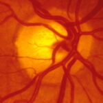
Here at the Optical Warehouse, our optometrists use various digital tools, including retinal photographs, to swiftly identify and address any emerging problems. By utilising a high-resolution camera to capture an image of the back of your eye, a retinal photograph enables your optometrist to closely and precisely examine your eyes. This examination can uncover symptoms such as damaged blood vessels caused by diabetes, age-related macular degeneration, and glaucoma — all potential culprits for vision loss if left untreated. Remarkably, retinal photographs can even unveil other serious health concerns beyond the realm of ocular health, such as brain tumours and strokes.
We understand the invaluable nature of your eyesight, which is why our highly qualified eye care professionals are dedicated to safeguarding your vision and overall eye health for the long haul. The process of retinal photography is rapid and painless, with immediate visualisation of the captured images.
Retinal Photography: An Overview
Retinal photography involves capturing a high-resolution digital image of the posterior (back) part of your eye. This image reveals crucial details, including your retina – a delicate layer of light-sensitive cells that captures light and images, along with your optic disk – a small focal point on the retina housing the optic nerve responsible for transmitting information to the brain and the intricate network of blood vessels.
Here at the Optical Warehouse, we prioritise comprehensive eye examinations, which encompass retinal photography. In Australia, it is recommended to undergo this examination every two years, or annually if you are 65 years or older. We securely store your digital images, allowing our optometry team to compare your retinal photographs from different visits. This facilitates a comprehensive analysis over time, enabling early detection and monitoring of any potential eye health concerns.
Different methods of retinal photography exist, each with its own distinct benefits. These methods include optical coherence tomography, fundus photography, and angiography. Your optometrist will carefully select the most appropriate imaging technique tailored to your specific eye and vision requirements, ensuring optimal accuracy and diagnostic capabilities.
What Can A Retinal Photograph Detect?
There are five common conditions that can be picked up by a retinal photograph, alongside others. These include:
Diabetic retinopathy
diabetic retinopathy arises when high blood sugar levels consistently damage the blood vessels in the retina, often affecting individuals with diabetes. In retinal photographs, diabetic retinopathy manifests through various symptoms, including swollen retinal veins, bleeding in the vitreous (the clear gel within the eye), the presence of new or abnormal blood vessels, and potential scar tissue or retinal detachment in advanced stages.
Hypertensive retinopathy
chronic hypertension, which is characterised by persistently high blood pressure, can lead to hypertensive retinopathy. Without proper treatment, this condition can progress and potentially cause vision loss. Indications of hypertensive retinopathy visible in retinal photographs encompass retinal exudates or fatty deposits, retinal bleeding, and the presence of white specks resembling cotton wool, which may suggest microstrokes.
Retinal tear and detachment
although this is more rare, retinal tears or complete detachment can occur, necessitating immediate medical attention as they can lead to vision loss. Retinal tears and detachments may be associated with various conditions, including age-related macular degeneration and eye injuries. Retinal photographs can exhibit signs of retinal damage or detachment such as grey discolouration of the retina, folded or detached areas of the retina, and twisted blood vessels.
Papilledema
papilledema refers to optic nerve swelling caused by increased pressure in the brain. It can be an indication of underlying conditions such as high blood pressure, eye or head injuries, encephalitis (brain inflammation), meningitis, stroke, or tumours in the eye or brain. Optometrists analyse retinal photographs to identify swollen optic nerves, bleeding near the optic nerve, and dilated or twisted blood vessels, which may suggest papilledema.
Optic atrophy
optic atrophy occurs when the optic nerve sustains damage, impeding the transmission of visual information from the eye to the brain. Multiple sclerosis, stroke, cranial arteritis, and brain tumours are among the medical conditions associated with optic atrophy, leading to gradual vision loss over time. Retinal photographs can disclose signs of optic atrophy, such as a pale white optic nerve, well-defined edges of the optic nerve, and normal veins alongside narrowed arteries.
Retinal Photography Available At The Optical Warehouse
It’s reassuring to know that many eye conditions and diseases can be detected in the very early stages before any noticeable symptoms occur, through retinal photography, which is included as part of our comprehensive eye examination with our qualified eye care professionals. As well as retinal photography, a comprehensive exam may also include:
• Going through your personal and family health history, detailing when any concerning symptoms began, medications you’re taking, work and environmental factors, and more
• Visual acuity measurements using reading charts to assess exactly how clearly each eye can see
• Tests of your eye health which may include depth perception, colour vision, peripheral (side) vision and how your pupils respond to light
• Assessments to measure which power of lens you require to correct near-sightedness, far-sightedness or astigmatism
• Eye focusing, eye teaming and eye movement activities to examine how well your eyes focus, move and work together
• Measurement of the pressure within the eye.
• Fluorescein angiography to detect any abnormal blood vessel growth.
After your exam is completed, your eye care professional will be able to discuss your diagnosis and treatment plan options with you to protect your eye health, best correct your vision, if needed, and help give you the freedom to do the things you love.
We’re proud to offer targeted care for a wide range of conditions. Every treatment plan is designed uniquely for your needs, preferences, and to help optimise your quality of life. Our optometrist will also be able to refer you to an ophthalmologist, or eye doctor specialising in surgery, for more complex treatment, if needed.
To book your retinal photography as part of your comprehensive eye exam with one of our eye care professionals, contact one of your local Optical Warehouse clinics across Queensland.


